Radiolucent PEEK body
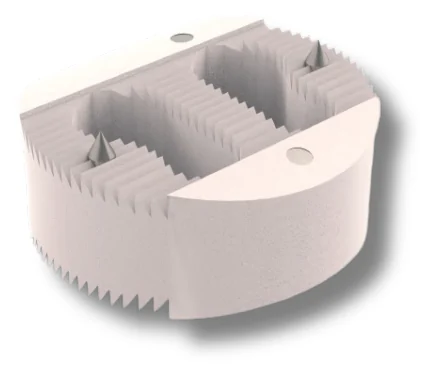
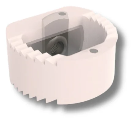
PEEK is radiolucent, so the cage is intended to leave the fusion area clear on imaging. Integrated tantalum markers show its position on fluoroscopy during insertion, which helps the surgeon judge depth and alignment before the holder is released.
Endplate-matched profile
The contoured profile follows the shape of the vertebral endplates to increase contact area.
Teeth and titanium pins
Serrations on the upper and lower surfaces and two titanium pins are designed to give initial fixation and to resist migration. A central opening is packed with graft before insertion.
Depth-set holder
The POLAR holder has a preset depth feature, so the cage is advanced to a defined position under fluoroscopy. Caspar pins and the Caspar retractor keep the segment distracted while the cage is tapped in.

 POLAR-BCervical bladed PEEK cage
 CARSTENTrabecular stand-alone cervical cage
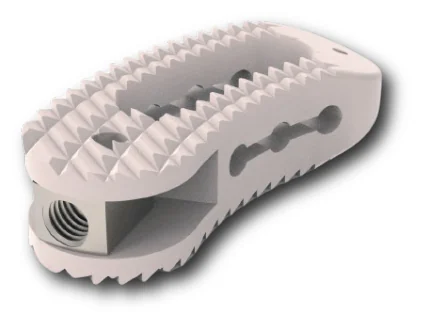 TWISTYTLIF PEEK cage
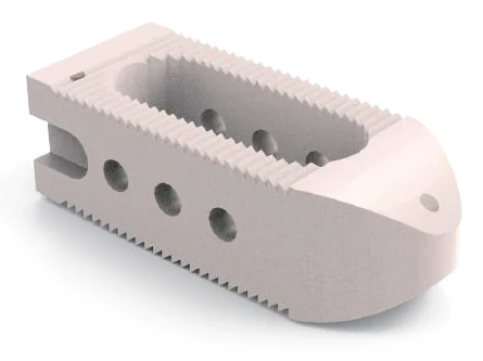 ALLIGATORPLIF PEEK cage
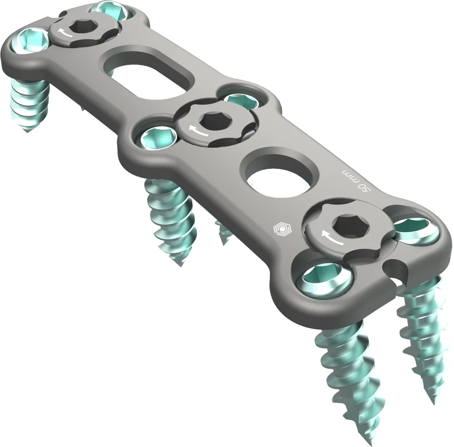 TRIMIAnterior cervical plate system
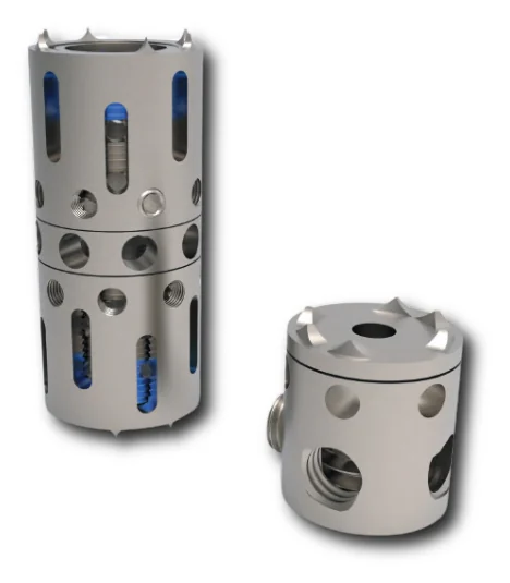 VERTELIFT-CAnterior cervical corpectomy cage
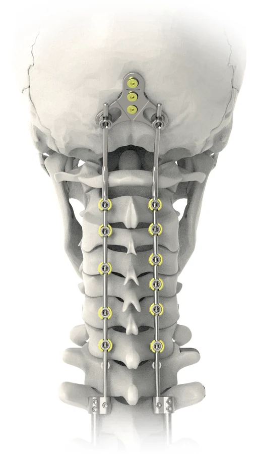 OXIS PLUSOccipitocervical fixation system
 D-FLEXTitanium cervical disc prosthesis